|
|
市場調査レポート
商品コード
1780320
顕微鏡の市場規模と予測 2021年~2031年、世界と地域のシェア、動向、成長機会分析レポート:技術別、エンドユーザー別、地域別Microscope Market Size and Forecast 2021-2031, Global and Regional Growth Opportunity Analysis Report Coverage: By Technology, End User, and Geography |
||||||

|
|||||||
|
|||||||
| 顕微鏡の市場規模と予測 2021年~2031年、世界と地域のシェア、動向、成長機会分析レポート:技術別、エンドユーザー別、地域別 |
|
出版日: 2025年07月03日
発行: The Insight Partners
ページ情報: 英文 184 Pages
納期: 即納可能
|
全表示
- 概要
- 図表
- 目次
顕微鏡市場規模は、2024年の26億5,000万米ドルから2031年には40億3,000万米ドルに達すると予測されています。2025~2031年のCAGRは6.20%と推定されます。市場開拓の主な要因としては、研究・ヘルスケア用途の増加、研究開発資金の増加などが挙げられます。さらに、顕微鏡にAIや遠隔技術を統合する動きが活発化しており、予測期間中に市場を押し上げる可能性が高いです。しかし、初期費用とメンテナンス費用が高いことが市場阻害要因の1つとなっています。
人工知能(AI)と遠隔技術の統合は、イメージング、データ分析、アクセシビリティの進歩につながる顕微鏡市場に革命をもたらしています。AIを搭載した顕微鏡システムは、画像解釈の精度、スピード、深度を向上させ、研究者や臨床医が複雑なサンプルから意味のある洞察をより効率的に引き出すことを可能にします。AIアルゴリズムは、細胞を自動的に識別・分類し、組織サンプルの異常を検出し、分子の相互作用をリアルタイムで定量化することができるため、人為的ミスを大幅に減らし、診断プロセスを加速します。デジタル技術とクラウド技術によって可能になった遠隔顕微鏡検査は、専門医が高解像度のイメージングと分析を遠隔で行うことを可能にし、AIの統合を補完します。この機能は特に遠隔医療において大きな変革をもたらし、遠隔診断により、十分なサービスを受けていない人々や地理的に孤立した人々にタイムリーで正確なヘルスケアサービスを提供することができます。また、リモート顕微鏡の使用は、世界中の複数の専門家が同時に顕微鏡画像をリアルタイムで閲覧・操作できるようにすることで、共同研究をサポートし、組織横断的なイノベーションと知識の共有を促進します。例えば、ツァイスのAI主導型デジタル顕微鏡プラットフォームは、自動画像解析とクラウドベースのデータ共有を組み合わせ、病理医が精度を高めたスライドをレビューできるようにしています。同様に、ライカマイクロシステムズは、同社の顕微鏡とシームレスに統合するAI支援ソフトウェアツールを開発し、従来の手作業による分析よりも迅速にがん細胞やその他の病理学的特徴を検出しています。さらに、AI一体型顕微鏡の発売は世界中で増加しています。例えば、2024年9月、MedPrime Technologies社は、インドのデジタル病理学に革命を起こす革新的なAI統合デジタル顕微鏡プラットフォームであるMICALYSを発売しました。診断精度を高め、ワークフローを合理化し、全体的な生産性を向上させる。これらのシステムは診断結果を改善するだけでなく、臨床検査室や研究室におけるワークフローの効率を最適化します。したがって、顕微鏡におけるAIと遠隔技術の統合の増加は、今後数年間の市場成長に寄与すると予想されます。
顕微鏡検査は医学教育やトレーニングにおいて非常に重要であり、学生や専門家が実践的なスキルを身につけ、人体解剖学や病理学をより深く理解することを可能にします。教育機関は、バーチャル顕微鏡や遠隔学習を可能にするインタラクティブなデジタル顕微鏡プラットフォームに投資しています。さらに、動物の健康状態をモニターし、環境汚染物質を検出し、農作物の収穫量を向上させるために、獣医学、環境科学、農業研究において顕微鏡は極めて重要です。例えば、電子顕微鏡は植物病理学で広く使用され、作物に影響を与えるウイルスや真菌を研究し、病気に強い植物品種の開発を促進しています。そのため、顕微鏡の技術的進歩と相まって、調査やヘルスケア用途の拡大が顕微鏡市場の成長を牽引しています。
さらに、政府、民間機関、組織は、ヘルスケア、バイオテクノロジー、ナノテクノロジー、材料科学などの科学研究開発により多くのリソースを割り当てています。このような資金の急増により、研究機関や大学は最先端の顕微鏡装置を調達することができます。例えば、米国の国立衛生研究所(NIH)は一貫して予算を増やし続け、2025年には480億米ドル以上に達します。この予算の大部分は、がん生物学から感染症研究に至るまで、顕微鏡イメージング技術に大きく依存する研究を支援しています。このような資金の流入により、研究室は従来の顕微鏡から、より高精度でより詳細なイメージング機能を提供する共焦点顕微鏡、電子顕微鏡、超解像顕微鏡などの高度なモデルへのアップグレードを促しています。
競合企業別分析では、製品ポートフォリオ(製品満足度、製品の特徴、入手可能性)、最近の市場動向(合併・買収、新製品の発売・強化、投資・資金調達、受賞、合意、提携・協力、認知、拡大)、競合状況のより良い意思決定と理解を助ける地域的プレゼンスに基づいて、試験管内肺モデル市場を評価・分類しています。本レポートでは、世界の試験管内肺モデル市場における主要ベンダーの最近の重要な動向と革新について深く調査しています。主な市場企業は、CARL ZEISS AG、Bruker Corporation、Leica Microsystems、Nikon Corporation、Thermo Fischer Scientific Inc.、Olympus Corporation、ACCU-SCOPE、Oxford Instruments Plc、Euromex Microscopen BV、Coxem Co.
顕微鏡市場は技術別に光学顕微鏡、電子顕微鏡、走査型プローブ顕微鏡、その他に区分されます。光学顕微鏡セグメントは2024年に顕微鏡市場で最大のシェアを占め、2025-2031年に大きなCAGRを記録すると予測されています。
エンドユーザーでは、顕微鏡市場は学術・研究機関、製薬・バイオ製薬企業、診断センター、その他に区分されます。製薬・バイオ製薬企業セグメントは、2024年に顕微鏡市場で最大のシェアを占め、2025-2031年に大きなCAGRを記録すると予測されています。大学、カレッジ、専用研究センターは、生物学、化学、材料科学、ナノテクノロジーなどの教育、基礎研究、実験研究に顕微鏡を幅広く活用しています。継続的な資金提供や助成金により、科学的知識や技術革新が促進されます。教室で使用される基本的な光学モデルから、最先端の研究ラボで使用される高度な電子顕微鏡や走査型プローブ顕微鏡まで、アカデミックな環境で使用される顕微鏡は多岐にわたる。光学顕微鏡は、依然として教育目的の最も一般的なツールであり、学生が細胞構造、微生物、組織サンプルを探索することを可能にしています。例えば、デジタル光学顕微鏡は、インタラクティブな学習体験を強化するために、カリキュラムに組み込まれることが多くなっています。一方、研究機関では、透過型電子顕微鏡(TEM)、走査型電子顕微鏡(SEM)、原子間力顕微鏡(AFM)、共焦点顕微鏡などのハイエンド技術に投資し、マイクロスケールやナノスケールでの詳細な構造、化学、物理分析を行うことが多いです。さらに、政府や民間団体は、科学インフラを支援するために多額の予算を割り当て続け、高度な顕微鏡システムのアップグレードや購入を推進しています。例えば、米国国立衛生研究所(NIH)や欧州研究会議(ERC)などのイニシアチブは、顕微鏡関連プロジェクトに積極的に資金を提供し、最先端の装置へのアクセスを容易にしています。
試験管内肺モデル市場で事業を展開する企業は、さまざまな有機的・無機的戦略を採用しています。有機的戦略には主に、製品上市と製品承認が含まれます。市場で見られる無機的成長戦略には、買収、提携、パートナーシップなどがあります。これらの成長戦略により、市場参入企業は事業を拡大し、地理的プレゼンスを高めるとともに、市場全体の成長に貢献することができます。さらに、買収や提携などの戦略は、顧客基盤を強化し、製品ポートフォリオを拡大するのに役立っています。試験管内肺モデル市場の主要企業による重要な発展の一部を以下に示します。
2024年10月、ZEISS社は、透過型電子顕微鏡(TEM)サンプルの完全自動調製に最適化された集束イオンビーム走査型電子顕微鏡(FIB-SEM)の新製品ZEISS Crossbeam 550 Samplefabを発売しました。
2024年6月、ライカマイクロシステムズは、脳神経外科用デジタル可視化顕微鏡ARveo 8の進化版を発表しました。ARveo 8は、3Dビューと拡張現実蛍光を適用することで、外科手術の可視化を強化します。
目次
第1章 イントロダクション
第2章 エグゼクティブサマリー
- アナリスト市場の展望
第3章 調査手法
- 2次調査
- 1次調査
- 仮説の策定
- マクロ経済要因分析
- ファンデーション数値の開発
- データの三角測量
- 国レベルのデータ
- 仮定と限界
第4章 顕微鏡市場情勢
- PEST分析
第5章 顕微鏡市場:主要市場力学
- 顕微鏡市場:主要市場力学
- 市場促進要因
- 調査とヘルスケア用途の増加
- 研究資金の急増
- 市場抑制要因
- 高い初期費用とメンテナンス費用
- 市場機会
- 市場企業による製品上市の増加
- 今後の動向
- 人工知能(AI)と遠隔技術の統合
- 促進要因と抑制要因の影響
第6章 顕微鏡市場:世界市場分析
- 顕微鏡市場の収益、2021-2031年
- 顕微鏡市場の予測分析
第7章 顕微鏡市場分析-技術別
- 光学顕微鏡
- 電子顕微鏡
- 走査型プローブ顕微鏡
- その他
第8章 顕微鏡市場分析-エンドユーザー別
- 学術・研究機関
- 製薬会社およびバイオ製薬会社
- 診断センター
- その他
第9章 顕微鏡市場-地域別市場分析
- 北米
- 米国
- カナダ
- メキシコ
- 欧州
- 英国
- ドイツ
- フランス
- イタリア
- スペイン
- その他欧州
- アジア太平洋
- 中国
- 日本
- インド
- オーストラリア
- 韓国
- アジア太平洋地域のその他諸国
- 中東・アフリカ
- 南アフリカ
- サウジアラビア
- アラブ首長国連邦
- その他中東とアフリカ
- 中南米
- ブラジル
- アルゼンチン
- その他中南米
第10章 競合情勢
- 主要企業によるヒートマップ分析
- 企業のポジショニングと集中度
第11章 顕微鏡の世界市場業界情勢
- 製品上市、製品承認
- 事業拡大、その他の戦略的開発
第12章 企業プロファイル
- Carl Zeiss AG
- Bruker Corp
- Leica Microsystems
- Nikon Corp
- Hitachi High-Tech Corp
- ACCU-SCOPE
- Thermo Fisher Scientific Inc
- Euromex Microscopen BV
- Oxford Instruments Plc
- COXEM Co.,Ltd
第13章 付録
List Of Tables
- Table 1. Microscope Market Segmentation
- Table 2. Microscope Market - Revenue, 2021-2024 (US$ Million)
- Table 3. Microscope Market - Revenue Forecast, 2025-2031 (US$ Million)
- Table 4. Microscope Market - Revenue, 2021-2024 (US$ Million) - by Technology
- Table 5. Microscope Market - Revenue Forecast, 2025-2031 (US$ Million) - by Technology
- Table 6. Microscope Market - Revenue, 2021-2024 (US$ Million) - by End User
- Table 7. Microscope Market - Revenue Forecast, 2025-2031 (US$ Million) - by End User
- Table 8. North America: Microscope Market - Revenue, 2021-2024 (US$ Million) - by Technology
- Table 9. North America: Microscope Market - Revenue Forecast, 2025-2031 (US$ Million) - by Technology
- Table 10. North America: Microscope Market - Revenue, 2021-2024 (US$ Million) - by End User
- Table 11. North America: Microscope Market - Revenue Forecast, 2025-2031 (US$ Million) - by End User
- Table 12. North America: Microscope Market - Revenue, 2021-2024 (US$ Million) - by Country
- Table 13. North America: Microscope Market - Revenue Forecast, 2025-2031 (US$ Million) - by Country
- Table 14. United States: Microscope Market - Revenue, 2021-2024 (US$ Million) - by Technology
- Table 15. United States: Microscope Market - Revenue Forecast, 2025-2031 (US$ Million) - by Technology
- Table 16. United States: Microscope Market - Revenue, 2021-2024 (US$ Million) - by End User
- Table 17. United States: Microscope Market - Revenue Forecast, 2025-2031 (US$ Million) - by End User
- Table 18. Canada: Microscope Market - Revenue, 2021-2024 (US$ Million) - by Technology
- Table 19. Canada: Microscope Market - Revenue Forecast, 2025-2031 (US$ Million) - by Technology
- Table 20. Canada: Microscope Market - Revenue, 2021-2024 (US$ Million) - by End User
- Table 21. Canada: Microscope Market - Revenue Forecast, 2025-2031 (US$ Million) - by End User
- Table 22. Mexico: Microscope Market - Revenue, 2021-2024 (US$ Million) - by Technology
- Table 23. Mexico: Microscope Market - Revenue Forecast, 2025-2031 (US$ Million) - by Technology
- Table 24. Mexico: Microscope Market - Revenue, 2021-2024 (US$ Million) - by End User
- Table 25. Mexico: Microscope Market - Revenue Forecast, 2025-2031 (US$ Million) - by End User
- Table 26. Europe: Microscope Market - Revenue, 2021-2024 (US$ Million) - by Technology
- Table 27. Europe: Microscope Market - Revenue Forecast, 2025-2031 (US$ Million) - by Technology
- Table 28. Europe: Microscope Market - Revenue, 2021-2024 (US$ Million) - by End User
- Table 29. Europe: Microscope Market - Revenue Forecast, 2025-2031 (US$ Million) - by End User
- Table 30. Europe: Microscope Market - Revenue, 2021-2024 (US$ Million) - by Country
- Table 31. Europe: Microscope Market - Revenue Forecast, 2025-2031 (US$ Million) - by Country
- Table 32. United Kingdom: Microscope Market - Revenue, 2021-2024 (US$ Million) - by Technology
- Table 33. United Kingdom: Microscope Market - Revenue Forecast, 2025-2031 (US$ Million) - by Technology
- Table 34. United Kingdom: Microscope Market - Revenue, 2021-2024 (US$ Million) - by End User
- Table 35. United Kingdom: Microscope Market - Revenue Forecast, 2025-2031 (US$ Million) - by End User
- Table 36. Germany: Microscope Market - Revenue, 2021-2024 (US$ Million) - by Technology
- Table 37. Germany: Microscope Market - Revenue Forecast, 2025-2031 (US$ Million) - by Technology
- Table 38. Germany: Microscope Market - Revenue, 2021-2024 (US$ Million) - by End User
- Table 39. Germany: Microscope Market - Revenue Forecast, 2025-2031 (US$ Million) - by End User
- Table 40. France: Microscope Market - Revenue, 2021-2024 (US$ Million) - by Technology
- Table 41. France: Microscope Market - Revenue Forecast, 2025-2031 (US$ Million) - by Technology
- Table 42. France: Microscope Market - Revenue, 2021-2024 (US$ Million) - by End User
- Table 43. France: Microscope Market - Revenue Forecast, 2025-2031 (US$ Million) - by End User
- Table 44. Italy: Microscope Market - Revenue, 2021-2024 (US$ Million) - by Technology
- Table 45. Italy: Microscope Market - Revenue Forecast, 2025-2031 (US$ Million) - by Technology
- Table 46. Italy: Microscope Market - Revenue, 2021-2024 (US$ Million) - by End User
- Table 47. Italy: Microscope Market - Revenue Forecast, 2025-2031 (US$ Million) - by End User
- Table 48. Spain: Microscope Market - Revenue, 2021-2024 (US$ Million) - by Technology
- Table 49. Spain: Microscope Market - Revenue Forecast, 2025-2031 (US$ Million) - by Technology
- Table 50. Spain: Microscope Market - Revenue, 2021-2024 (US$ Million) - by End User
- Table 51. Spain: Microscope Market - Revenue Forecast, 2025-2031 (US$ Million) - by End User
- Table 52. Rest of Europe: Microscope Market - Revenue, 2021-2024 (US$ Million) - by Technology
- Table 53. Rest of Europe: Microscope Market - Revenue Forecast, 2025-2031 (US$ Million) - by Technology
- Table 54. Rest of Europe: Microscope Market - Revenue, 2021-2024 (US$ Million) - by End User
- Table 55. Rest of Europe: Microscope Market - Revenue Forecast, 2025-2031 (US$ Million) - by End User
- Table 56. Asia Pacific: Microscope Market - Revenue, 2021-2024 (US$ Million) - by Technology
- Table 57. Asia Pacific: Microscope Market - Revenue Forecast, 2025-2031 (US$ Million) - by Technology
- Table 58. Asia Pacific: Microscope Market - Revenue, 2021-2024 (US$ Million) - by End User
- Table 59. Asia Pacific: Microscope Market - Revenue Forecast, 2025-2031 (US$ Million) - by End User
- Table 60. Asia Pacific: Microscope Market - Revenue, 2021-2024 (US$ Million) - by Country
- Table 61. Asia Pacific: Microscope Market - Revenue Forecast, 2025-2031 (US$ Million) - by Country
- Table 62. China: Microscope Market - Revenue, 2021-2024 (US$ Million) - by Technology
- Table 63. China: Microscope Market - Revenue Forecast, 2025-2031 (US$ Million) - by Technology
- Table 64. China: Microscope Market - Revenue, 2021-2024 (US$ Million) - by End User
- Table 65. China: Microscope Market - Revenue Forecast, 2025-2031 (US$ Million) - by End User
- Table 66. Japan: Microscope Market - Revenue, 2021-2024 (US$ Million) - by Technology
- Table 67. Japan: Microscope Market - Revenue Forecast, 2025-2031 (US$ Million) - by Technology
- Table 68. Japan: Microscope Market - Revenue, 2021-2024 (US$ Million) - by End User
- Table 69. Japan: Microscope Market - Revenue Forecast, 2025-2031 (US$ Million) - by End User
- Table 70. India: Microscope Market - Revenue, 2021-2024 (US$ Million) - by Technology
- Table 71. India: Microscope Market - Revenue Forecast, 2025-2031 (US$ Million) - by Technology
- Table 72. India: Microscope Market - Revenue, 2021-2024 (US$ Million) - by End User
- Table 73. India: Microscope Market - Revenue Forecast, 2025-2031 (US$ Million) - by End User
- Table 74. Australia: Microscope Market - Revenue, 2021-2024 (US$ Million) - by Technology
- Table 75. Australia: Microscope Market - Revenue Forecast, 2025-2031 (US$ Million) - by Technology
- Table 76. Australia: Microscope Market - Revenue, 2021-2024 (US$ Million) - by End User
- Table 77. Australia: Microscope Market - Revenue Forecast, 2025-2031 (US$ Million) - by End User
- Table 78. South Korea: Microscope Market - Revenue, 2021-2024 (US$ Million) - by Technology
- Table 79. South Korea: Microscope Market - Revenue Forecast, 2025-2031 (US$ Million) - by Technology
- Table 80. South Korea: Microscope Market - Revenue, 2021-2024 (US$ Million) - by End User
- Table 81. South Korea: Microscope Market - Revenue Forecast, 2025-2031 (US$ Million) - by End User
- Table 82. Rest of APAC: Microscope Market - Revenue, 2021-2024 (US$ Million) - by Technology
- Table 83. Rest of APAC: Microscope Market - Revenue Forecast, 2025-2031 (US$ Million) - by Technology
- Table 84. Rest of APAC: Microscope Market - Revenue, 2021-2024 (US$ Million) - by End User
- Table 85. Rest of APAC: Microscope Market - Revenue Forecast, 2025-2031 (US$ Million) - by End User
- Table 86. Middle East and Africa: Microscope Market - Revenue, 2021-2024 (US$ Million) - by Technology
- Table 87. Middle East and Africa: Microscope Market - Revenue Forecast, 2025-2031 (US$ Million) - by Technology
- Table 88. Middle East and Africa: Microscope Market - Revenue, 2021-2024 (US$ Million) - by End User
- Table 89. Middle East and Africa: Microscope Market - Revenue Forecast, 2025-2031 (US$ Million) - by End User
- Table 90. Middle East and Africa: Microscope Market - Revenue, 2021-2024 (US$ Million) - by Country
- Table 91. Middle East and Africa: Microscope Market - Revenue Forecast, 2025-2031 (US$ Million) - by Country
- Table 92. South Africa: Microscope Market - Revenue, 2021-2024 (US$ Million) - by Technology
- Table 93. South Africa: Microscope Market - Revenue Forecast, 2025-2031 (US$ Million) - by Technology
- Table 94. South Africa: Microscope Market - Revenue, 2021-2024 (US$ Million) - by End User
- Table 95. South Africa: Microscope Market - Revenue Forecast, 2025-2031 (US$ Million) - by End User
- Table 96. Saudi Arabia: Microscope Market - Revenue, 2021-2024 (US$ Million) - by Technology
- Table 97. Saudi Arabia: Microscope Market - Revenue Forecast, 2025-2031 (US$ Million) - by Technology
- Table 98. Saudi Arabia: Microscope Market - Revenue, 2021-2024 (US$ Million) - by End User
- Table 99. Saudi Arabia: Microscope Market - Revenue Forecast, 2025-2031 (US$ Million) - by End User
- Table 100. United Arab Emirates: Microscope Market - Revenue, 2021-2024 (US$ Million) - by Technology
- Table 101. United Arab Emirates: Microscope Market - Revenue Forecast, 2025-2031 (US$ Million) - by Technology
- Table 102. United Arab Emirates: Microscope Market - Revenue, 2021-2024 (US$ Million) - by End User
- Table 103. United Arab Emirates: Microscope Market - Revenue Forecast, 2025-2031 (US$ Million) - by End User
- Table 104. Rest of Middle East and Africa: Microscope Market - Revenue, 2021-2024 (US$ Million) - by Technology
- Table 105. Rest of Middle East and Africa: Microscope Market - Revenue Forecast, 2025-2031 (US$ Million) - by Technology
- Table 106. Rest of Middle East and Africa: Microscope Market - Revenue, 2021-2024 (US$ Million) - by End User
- Table 107. Rest of Middle East and Africa: Microscope Market - Revenue Forecast, 2025-2031 (US$ Million) - by End User
- Table 108. South and Central America: Microscope Market - Revenue, 2021-2024 (US$ Million) - by Technology
- Table 109. South and Central America: Microscope Market - Revenue Forecast, 2025-2031 (US$ Million) - by Technology
- Table 110. South and Central America: Microscope Market - Revenue, 2021-2024 (US$ Million) - by End User
- Table 111. South and Central America: Microscope Market - Revenue Forecast, 2025-2031 (US$ Million) - by End User
- Table 112. South and Central America: Microscope Market - Revenue, 2021-2024 (US$ Million) - by Country
- Table 113. South and Central America: Microscope Market - Revenue Forecast, 2025-2031 (US$ Million) - by Country
- Table 114. Brazil: Microscope Market - Revenue, 2021-2024 (US$ Million) - by Technology
- Table 115. Brazil: Microscope Market - Revenue Forecast, 2025-2031 (US$ Million) - by Technology
- Table 116. Brazil: Microscope Market - Revenue, 2021-2024 (US$ Million) - by End User
- Table 117. Brazil: Microscope Market - Revenue Forecast, 2025-2031 (US$ Million) - by End User
- Table 118. Argentina: Microscope Market - Revenue, 2021-2024 (US$ Million) - by Technology
- Table 119. Argentina: Microscope Market - Revenue Forecast, 2025-2031 (US$ Million) - by Technology
- Table 120. Argentina: Microscope Market - Revenue, 2021-2024 (US$ Million) - by End User
- Table 121. Argentina: Microscope Market - Revenue Forecast, 2025-2031 (US$ Million) - by End User
- Table 122. Rest of South and Central America: Microscope Market - Revenue, 2021-2024 (US$ Million) - by Technology
- Table 123. Rest of South and Central America: Microscope Market - Revenue Forecast, 2025-2031 (US$ Million) - by Technology
- Table 124. Rest of South and Central America: Microscope Market - Revenue, 2021-2024 (US$ Million) - by End User
- Table 125. Rest of South and Central America: Microscope Market - Revenue Forecast, 2025-2031 (US$ Million) - by End User
- Table 126. Heat Map Analysis by Key Players
- Table 127. Glossary of Terms
List Of Figures
- Figure 1. Microscope Market Segmentation, by Geography
- Figure 2. PEST Analysis
- Figure 3. Impact Analysis of Drivers and Restraints
- Figure 4. Microscope Market Revenue (US$ Million), 2021-2031
- Figure 5. Microscope Market Share (%) - by Technology (2024 and 2031)
- Figure 6. Optical Microscope: Microscope Market - Revenue and Forecast to 2031 (US$ Million)
- Figure 7. Electron Microscope: Microscope Market - Revenue and Forecast to 2031 (US$ Million)
- Figure 8. Scanning Probe Microscope: Microscope Market - Revenue and Forecast to 2031 (US$ Million)
- Figure 9. Others: Microscope Market - Revenue and Forecast to 2031 (US$ Million)
- Figure 10. Microscope Market Share (%) - by End User (2024 and 2031)
- Figure 11. Academics and Research Institutes: Microscope Market - Revenue and Forecast to 2031 (US$ Million)
- Figure 12. Pharmaceuticals and Biopharmaceutical Companies: Microscope Market - Revenue and Forecast to 2031 (US$ Million)
- Figure 13. Diagnostic Centers: Microscope Market - Revenue and Forecast to 2031 (US$ Million)
- Figure 14. Others: Microscope Market - Revenue and Forecast to 2031 (US$ Million)
- Figure 15. Microscope Market Breakdown by Region, 2024 and 2031 (%)
- Figure 16. North America: Microscope Market - Revenue, 2021-2031 (US$ Million)
- Figure 17. North America: Microscope Market Breakdown, by Technology (2024 and 2031)
- Figure 18. North America: Microscope Market Breakdown, by End User (2024 and 2031)
- Figure 19. North America: Microscope Market Breakdown, by Key Countries, 2024 and 2031 (%)
- Figure 20. United States: Microscope Market - Revenue and Forecast, 2021-2031 (US$ Million)
- Figure 21. Canada: Microscope Market - Revenue and Forecast, 2021-2031 (US$ Million)
- Figure 22. Mexico: Microscope Market - Revenue and Forecast, 2021-2031 (US$ Million)
- Figure 23. Europe: Microscope Market - Revenue, 2021-2031 (US$ Million)
- Figure 24. Europe: Microscope Market Breakdown, by Technology (2024 and 2031)
- Figure 25. Europe: Microscope Market Breakdown, by End User (2024 and 2031)
- Figure 26. Europe: Microscope Market Breakdown, by Key Countries, 2024 and 2031 (%)
- Figure 27. United Kingdom: Microscope Market - Revenue and Forecast, 2021-2031 (US$ Million)
- Figure 28. Germany: Microscope Market - Revenue and Forecast, 2021-2031 (US$ Million)
- Figure 29. France: Microscope Market - Revenue and Forecast, 2021-2031 (US$ Million)
- Figure 30. Italy: Microscope Market - Revenue and Forecast, 2021-2031 (US$ Million)
- Figure 31. Spain: Microscope Market - Revenue and Forecast, 2021-2031 (US$ Million)
- Figure 32. Rest of Europe: Microscope Market - Revenue and Forecast, 2021-2031 (US$ Million)
- Figure 33. Asia Pacific: Microscope Market - Revenue, 2021-2031 (US$ Million)
- Figure 34. Asia Pacific: Microscope Market Breakdown, by Technology (2024 and 2031)
- Figure 35. Asia Pacific: Microscope Market Breakdown, by End User (2024 and 2031)
- Figure 36. Asia Pacific: Microscope Market Breakdown, by Key Countries, 2024 and 2031 (%)
- Figure 37. China: Microscope Market - Revenue and Forecast, 2021-2031 (US$ Million)
- Figure 38. Japan: Microscope Market - Revenue and Forecast, 2021-2031 (US$ Million)
- Figure 39. India: Microscope Market - Revenue and Forecast, 2021-2031 (US$ Million)
- Figure 40. Australia: Microscope Market - Revenue and Forecast, 2021-2031 (US$ Million)
- Figure 41. South Korea: Microscope Market - Revenue and Forecast, 2021-2031 (US$ Million)
- Figure 42. Rest of APAC: Microscope Market - Revenue and Forecast, 2021-2031 (US$ Million)
- Figure 43. Middle East and Africa: Microscope Market - Revenue, 2021-2031 (US$ Million)
- Figure 44. Middle East and Africa: Microscope Market Breakdown, by Technology (2024 and 2031)
- Figure 45. Middle East and Africa: Microscope Market Breakdown, by End User (2024 and 2031)
- Figure 46. Middle East and Africa: Microscope Market Breakdown, by Key Countries, 2024 and 2031 (%)
- Figure 47. South Africa: Microscope Market - Revenue and Forecast, 2021-2031 (US$ Million)
- Figure 48. Saudi Arabia: Microscope Market - Revenue and Forecast, 2021-2031 (US$ Million)
- Figure 49. United Arab Emirates: Microscope Market - Revenue and Forecast, 2021-2031 (US$ Million)
- Figure 50. Rest of Middle East and Africa: Microscope Market - Revenue and Forecast, 2021-2031 (US$ Million)
- Figure 51. South and Central America: Microscope Market - Revenue, 2021-2031 (US$ Million)
- Figure 52. South and Central America: Microscope Market Breakdown, by Technology (2024 and 2031)
- Figure 53. South and Central America: Microscope Market Breakdown, by End User (2024 and 2031)
- Figure 54. South and Central America: Microscope Market Breakdown, by Key Countries, 2024 and 2031 (%)
- Figure 55. Brazil: Microscope Market - Revenue and Forecast, 2021-2031 (US$ Million)
- Figure 56. Argentina: Microscope Market - Revenue and Forecast, 2021-2031 (US$ Million)
- Figure 57. Rest of South and Central America: Microscope Market - Revenue and Forecast, 2021-2031 (US$ Million)
- Figure 58. Company Positioning & Concentration
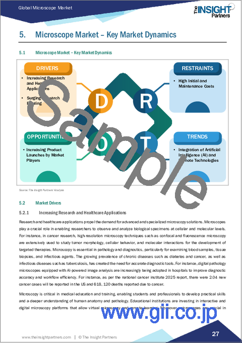
The microscope market size is projected to reach US$ 4.03 billion by 2031 from US$ 2.65 billion in 2024. The market is estimated to register a CAGR of 6.20% during 2025-2031. Major factors driving the market growth include an increasing applications of research and healthcare, and the growing funding in research and development. Further, increasing integration of AI and remote technologies in the microscope is likely to boost the market during the forecast period. However, high initial and maintenance costs are among the market deterrents.
The integration of Artificial Intelligence (AI) and remote technologies is revolutionizing the microscope market leading to advancements in imaging, data analysis, and accessibility. AI-powered microscopy systems enhance the precision, speed, and depth of image interpretation, enabling researchers and clinicians to extract meaningful insights from complex samples with greater efficiency. AI algorithms can automatically identify and classify cells, detect abnormalities in tissue samples, and quantify molecular interactions in real time, which significantly reduces human error and accelerates diagnostic processes. Remote microscopy, enabled by digital and cloud technologies, complements AI integration by allowing specialists to conduct high-resolution imaging and analysis remotely. This capability is particularly transformative in telemedicine, where remote diagnostics can provide timely and accurate healthcare services to underserved or geographically isolated populations. The use of remote microscopy also supports collaborative research by enabling multiple experts across the globe to simultaneously view and manipulate microscopic images in real time, fostering cross-institutional innovation and knowledge sharing. For instance, Zeiss's AI-driven digital microscopy platform combines automated image analysis with cloud-based data sharing, allowing pathologists to review slides with enhanced accuracy. Similarly, Leica Microsystems has developed AI-assisted software tools that integrate seamlessly with their microscopes to detect cancer cells and other pathological features more rapidly than traditional manual analysis. Moreover, the launch of AI-integrated microscopes is increasing across the world. For instance, in September 2024, MedPrime Technologies launched MICALYS, which is an innovative AI-integrated digital microscopy platform that is set to revolutionize digital pathology in India. It elevates diagnostic precision, streamlines workflows, and boosts overall productivity. These systems not only improve diagnostic outcomes but also optimize workflow efficiencies in clinical and research laboratories. Therefore, increasing integration of AI and remote technologies in the microscope is expected to contribute the market growth in the coming years.
Microscopy is critical in medical education and training, enabling students and professionals to develop practical skills and a deeper understanding of human anatomy and pathology. Educational institutions are investing in interactive and digital microscopy platforms that allow virtual microscopy and remote learning. Moreover, microscopes are crucial in veterinary medicine, environmental science, and agricultural research to monitor animal health, detect environmental contaminants, and improve crop yields. For instance, electron microscopy is widely used in plant pathology to study viruses and fungi affecting crops, facilitating the development of disease-resistant plant varieties. Therefore, the expanding scope of research and healthcare applications, coupled with technological advancements in microscopy, drives the growth of the microscope market.
Moreover, governments, private institutions, and organizations are allocating more resources toward scientific research and development in healthcare, biotechnology, nanotechnology, and materials science, among others. This surge in funding enables research institutions and universities to procure state-of-the-art microscopy equipment. For instance, the National Institutes of Health (NIH) in the US has consistently increased its budget, reaching over US$ 48 billion in 2025. A significant portion of this budget supports research that relies heavily on microscopic imaging technologies, from cancer biology to infectious disease studies. This influx of funds encourages laboratories to upgrade from conventional microscopes to advanced models such as confocal, electron, and super-resolution microscopes that offer higher precision and more detailed imaging capabilities
The comparative company analysis evaluates and categorizes the in vitro lung models market based on product portfolio (product satisfaction, product features, and availability), recent market developments (merger & acquisition, new product launch & enhancement, investment & funding, award, agreement, collaboration, & partnership, recognition, and expansion), and geographic presence that aids better decision-making and understanding of the competitive landscape. The report profoundly explores the recent significant developments and innovations by the leading vendors in the global in vitro lung models market. The key market players are CARL ZEISS AG; Bruker Corporation; Leica Microsystems; Nikon Corporation; Thermo Fischer Scientific Inc.; Olympus Corporation; ACCU-SCOPE; Oxford Instruments Plc; Euromex Microscopen BV; Coxem Co.,Ltd; and Hitachi High-Tech Corp.
Based on technology, the microscope market is segmented into optical microscope, electron microscope, scanning probe microscope, and others. The optical microscope segment held the largest share of the microscope market in 2024, and it is expected to register a significant CAGR during 2025-2031.
In terms of end user, the microscope market is segmented into academics and research institutes, pharmaceuticals and biopharmaceutical companies, diagnostic centers, and others. The pharmaceuticals and biopharmaceutical companies segment held the largest share of the microscope market in 2024, and it is expected to register a significant CAGR during 2025-2031. Universities, colleges, and dedicated research centers utilize microscopes extensively for education, fundamental research, and experimental studies in biology, chemistry, materials science, nanotechnology, and others. Continuous funding and grants enhance scientific knowledge and innovations. Microscopes in academic settings range from basic optical models used in classrooms to sophisticated electron and scanning probe microscopes employed in cutting-edge research labs. Optical microscopes remain the most common tool for teaching purposes, enabling students to explore cell structures, microorganisms, and tissue samples. For instance, digital optical microscopes are increasingly integrated into curricula to enhance interactive learning experiences. Meanwhile, research institutes often invest in high-end technologies such as transmission electron microscopes (TEM), scanning electron microscopes (SEM), atomic force microscopes (AFM), and confocal microscopes to conduct detailed structural, chemical, and physical analyses at the micro and nanoscale. Additionally, governments and private organizations continue to allocate significant budgets to support scientific infrastructure, driving upgrades and purchases of advanced microscopy systems. For example, initiatives such as the US National Institutes of Health (NIH) and the European Research Council (ERC) actively fund microscopy-related projects, facilitating access to state-of-the-art equipment.
Various organic and inorganic strategies are adopted by companies operating in the in vitro lung models market. The organic strategies mainly include product launches and product approvals. Inorganic growth strategies witnessed in the market are acquisitions, collaboration, and partnerships. These growth strategies allow the market players to expand their businesses and enhance their geographic presence, along with contributing to the overall market growth. Furthermore, strategies such as acquisitions and partnerships helped strengthen their customer base and extend their product portfolios. A few of the significant developments by key players in the in vitro lung models market are listed below.
In October 2024, ZEISS launched new ZEISS Crossbeam 550 Samplefab, a focused ion beam scanning electron microscope (FIB-SEM) optimized for fully automated preparation of transmission electron microscopy (TEM) samples.
In June 2024, Leica Microsystems introduced an evolved version of its ARveo 8 digital visualization microscope for neurosurgery. The ARveo 8 enhances surgical visualization by applying a 3D view and augmented reality fluorescence.
Table Of Contents
1. Introduction
- 1.1 Report Guidance
- 1.2 Market Segmentation
2. Executive Summary
- 2.1 Analyst Market Outlook
3. Research Methodology
- 3.1 Secondary Research
- 3.2 Primary Research
- 3.2.1 Hypothesis formulation:
- 3.2.2 Macro-economic factor analysis:
- 3.2.3 Developing base number:
- 3.2.4 Data Triangulation:
- 3.2.5 Country level data:
- 3.3 Assumptions and Limitations
4. Microscope Market Landscape
- 4.1 Overview
- 4.2 PEST Analysis
5. Microscope Market - Key Market Dynamics
- 5.1 Microscope Market - Key Market Dynamics
- 5.2 Market Drivers
- 5.2.1 Increasing Research and Healthcare Applications
- 5.2.2 Surging Research Funding
- 5.3 Market Restraints
- 5.3.1 High Initial and Maintenance Costs
- 5.4 Market Opportunities
- 5.4.1 Increasing Product Launches by Market Players
- 5.5 Future Trends
- 5.5.1 Integration of Artificial Intelligence (AI) and Remote Technologies
- 5.6 Impact of Drivers and Restraints:
6. Microscope Market - Global Market Analysis
- 6.1 Microscope Market Revenue (US$ Million), 2021-2031
- 6.2 Microscope Market Forecast Analysis
7. Microscope Market Analysis - by Technology
- 7.1 Optical Microscope
- 7.1.1 Overview
- 7.1.2 Optical Microscope: Microscope Market - Revenue and Forecast to 2031 (US$ Million)
- 7.2 Electron Microscope
- 7.2.1 Overview
- 7.2.2 Electron Microscope: Microscope Market - Revenue and Forecast to 2031 (US$ Million)
- 7.3 Scanning Probe Microscope
- 7.3.1 Overview
- 7.3.2 Scanning Probe Microscope: Microscope Market - Revenue and Forecast to 2031 (US$ Million)
- 7.4 Others
- 7.4.1 Overview
- 7.4.2 Others: Microscope Market - Revenue and Forecast to 2031 (US$ Million)
8. Microscope Market Analysis - by End User
- 8.1 Academics and Research Institutes
- 8.1.1 Overview
- 8.1.2 Academics and Research Institutes: Microscope Market - Revenue and Forecast to 2031 (US$ Million)
- 8.2 Pharmaceuticals and Biopharmaceutical Companies
- 8.2.1 Overview
- 8.2.2 Pharmaceuticals and Biopharmaceutical Companies: Microscope Market - Revenue and Forecast to 2031 (US$ Million)
- 8.3 Diagnostic Centers
- 8.3.1 Overview
- 8.3.2 Diagnostic Centers: Microscope Market - Revenue and Forecast to 2031 (US$ Million)
- 8.4 Others
- 8.4.1 Overview
- 8.4.2 Others: Microscope Market - Revenue and Forecast to 2031 (US$ Million)
9. Microscope Market - Geographical Analysis
- 9.1 Overview
- 9.2 North America
- 9.2.1 North America Microscope Market Overview
- 9.2.2 North America: Microscope Market - Revenue, 2021-2031 (US$ Million)
- 9.2.3 North America: Microscope Market Breakdown, by Technology
- 9.2.3.1 North America: Microscope Market - Revenue and Forecast Analysis - by Technology
- 9.2.4 North America: Microscope Market Breakdown, by End User
- 9.2.4.1 North America: Microscope Market - Revenue and Forecast Analysis - by End User
- 9.2.5 North America: Microscope Market - Revenue and Forecast Analysis - by Country
- 9.2.5.1 North America: Microscope Market - Revenue and Forecast Analysis - by Country
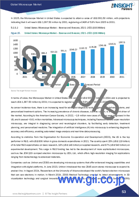
- 9.2.5.2 United States: Microscope Market - Revenue and Forecast, 2021-2031 (US$ Million)
- 9.2.5.2.1 United States: Microscope Market Breakdown, by Technology
- 9.2.5.2.2 United States: Microscope Market Breakdown, by End User
- 9.2.5.3 Canada: Microscope Market - Revenue and Forecast, 2021-2031 (US$ Million)
- 9.2.5.3.1 Canada: Microscope Market Breakdown, by Technology
- 9.2.5.3.2 Canada: Microscope Market Breakdown, by End User
- 9.2.5.4 Mexico: Microscope Market - Revenue and Forecast, 2021-2031 (US$ Million)
- 9.2.5.4.1 Mexico: Microscope Market Breakdown, by Technology
- 9.2.5.4.2 Mexico: Microscope Market Breakdown, by End User
- 9.3 Europe
- 9.3.1 Europe Microscope Market Overview
- 9.3.2 Europe: Microscope Market - Revenue, 2021-2031 (US$ Million)
- 9.3.3 Europe: Microscope Market Breakdown, by Technology
- 9.3.3.1 Europe: Microscope Market - Revenue and Forecast Analysis - by Technology
- 9.3.4 Europe: Microscope Market Breakdown, by End User
- 9.3.4.1 Europe: Microscope Market - Revenue and Forecast Analysis - by End User
- 9.3.5 Europe: Microscope Market - Revenue and Forecast Analysis - by Country
- 9.3.5.1 Europe: Microscope Market - Revenue and Forecast Analysis - by Country
- 9.3.5.2 United Kingdom: Microscope Market - Revenue and Forecast, 2021-2031 (US$ Million)
- 9.3.5.2.1 United Kingdom: Microscope Market Breakdown, by Technology
- 9.3.5.2.2 United Kingdom: Microscope Market Breakdown, by End User
- 9.3.5.3 Germany: Microscope Market - Revenue and Forecast, 2021-2031 (US$ Million)
- 9.3.5.3.1 Germany: Microscope Market Breakdown, by Technology
- 9.3.5.3.2 Germany: Microscope Market Breakdown, by End User
- 9.3.5.4 France: Microscope Market - Revenue and Forecast, 2021-2031 (US$ Million)
- 9.3.5.4.1 France: Microscope Market Breakdown, by Technology
- 9.3.5.4.2 France: Microscope Market Breakdown, by End User
- 9.3.5.5 Italy: Microscope Market - Revenue and Forecast, 2021-2031 (US$ Million)
- 9.3.5.5.1 Italy: Microscope Market Breakdown, by Technology
- 9.3.5.5.2 Italy: Microscope Market Breakdown, by End User
- 9.3.5.6 Spain: Microscope Market - Revenue and Forecast, 2021-2031 (US$ Million)
- 9.3.5.6.1 Spain: Microscope Market Breakdown, by Technology
- 9.3.5.6.2 Spain: Microscope Market Breakdown, by End User
- 9.3.5.7 Rest of Europe: Microscope Market - Revenue and Forecast, 2021-2031 (US$ Million)
- 9.3.5.7.1 Rest of Europe: Microscope Market Breakdown, by Technology
- 9.3.5.7.2 Rest of Europe: Microscope Market Breakdown, by End User
- 9.4 Asia Pacific
- 9.4.1 Asia Pacific Microscope Market Overview
- 9.4.2 Asia Pacific: Microscope Market - Revenue, 2021-2031 (US$ Million)
- 9.4.3 Asia Pacific: Microscope Market Breakdown, by Technology
- 9.4.3.1 Asia Pacific: Microscope Market - Revenue and Forecast Analysis - by Technology
- 9.4.4 Asia Pacific: Microscope Market Breakdown, by End User
- 9.4.4.1 Asia Pacific: Microscope Market - Revenue and Forecast Analysis - by End User
- 9.4.5 Asia Pacific: Microscope Market - Revenue and Forecast Analysis - by Country
- 9.4.5.1 Asia Pacific: Microscope Market - Revenue and Forecast Analysis - by Country
- 9.4.5.2 China: Microscope Market - Revenue and Forecast, 2021-2031 (US$ Million)
- 9.4.5.2.1 China: Microscope Market Breakdown, by Technology
- 9.4.5.2.2 China: Microscope Market Breakdown, by End User
- 9.4.5.3 Japan: Microscope Market - Revenue and Forecast, 2021-2031 (US$ Million)
- 9.4.5.3.1 Japan: Microscope Market Breakdown, by Technology
- 9.4.5.3.2 Japan: Microscope Market Breakdown, by End User
- 9.4.5.4 India: Microscope Market - Revenue and Forecast, 2021-2031 (US$ Million)
- 9.4.5.4.1 India: Microscope Market Breakdown, by Technology
- 9.4.5.4.2 India: Microscope Market Breakdown, by End User
- 9.4.5.5 Australia: Microscope Market - Revenue and Forecast, 2021-2031 (US$ Million)
- 9.4.5.5.1 Australia: Microscope Market Breakdown, by Technology
- 9.4.5.5.2 Australia: Microscope Market Breakdown, by End User
- 9.4.5.6 South Korea: Microscope Market - Revenue and Forecast, 2021-2031 (US$ Million)
- 9.4.5.6.1 South Korea: Microscope Market Breakdown, by Technology
- 9.4.5.6.2 South Korea: Microscope Market Breakdown, by End User
- 9.4.5.7 Rest of APAC: Microscope Market - Revenue and Forecast, 2021-2031 (US$ Million)
- 9.4.5.7.1 Rest of APAC: Microscope Market Breakdown, by Technology
- 9.4.5.7.2 Rest of APAC: Microscope Market Breakdown, by End User
- 9.5 Middle East and Africa
- 9.5.1 Middle East and Africa Microscope Market Overview
- 9.5.2 Middle East and Africa: Microscope Market - Revenue, 2021-2031 (US$ Million)
- 9.5.3 Middle East and Africa: Microscope Market Breakdown, by Technology
- 9.5.3.1 Middle East and Africa: Microscope Market - Revenue and Forecast Analysis - by Technology
- 9.5.4 Middle East and Africa: Microscope Market Breakdown, by End User
- 9.5.4.1 Middle East and Africa: Microscope Market - Revenue and Forecast Analysis - by End User
- 9.5.5 Middle East and Africa: Microscope Market - Revenue and Forecast Analysis - by Country
- 9.5.5.1 Middle East and Africa: Microscope Market - Revenue and Forecast Analysis - by Country
- 9.5.5.2 South Africa: Microscope Market - Revenue and Forecast, 2021-2031 (US$ Million)
- 9.5.5.2.1 South Africa: Microscope Market Breakdown, by Technology
- 9.5.5.2.2 South Africa: Microscope Market Breakdown, by End User
- 9.5.5.3 Saudi Arabia: Microscope Market - Revenue and Forecast, 2021-2031 (US$ Million)
- 9.5.5.3.1 Saudi Arabia: Microscope Market Breakdown, by Technology
- 9.5.5.3.2 Saudi Arabia: Microscope Market Breakdown, by End User
- 9.5.5.4 United Arab Emirates: Microscope Market - Revenue and Forecast, 2021-2031 (US$ Million)
- 9.5.5.4.1 United Arab Emirates: Microscope Market Breakdown, by Technology
- 9.5.5.4.2 United Arab Emirates: Microscope Market Breakdown, by End User
- 9.5.5.5 Rest of Middle East and Africa: Microscope Market - Revenue and Forecast, 2021-2031 (US$ Million)
- 9.5.5.5.1 Rest of Middle East and Africa: Microscope Market Breakdown, by Technology
- 9.5.5.5.2 Rest of Middle East and Africa: Microscope Market Breakdown, by End User
- 9.6 South and Central America
- 9.6.1 South and Central America Microscope Market Overview
- 9.6.2 South and Central America: Microscope Market - Revenue, 2021-2031 (US$ Million)
- 9.6.3 South and Central America: Microscope Market Breakdown, by Technology
- 9.6.3.1 South and Central America: Microscope Market - Revenue and Forecast Analysis - by Technology
- 9.6.4 South and Central America: Microscope Market Breakdown, by End User
- 9.6.4.1 South and Central America: Microscope Market - Revenue and Forecast Analysis - by End User
- 9.6.5 South and Central America: Microscope Market - Revenue and Forecast Analysis - by Country
- 9.6.5.1 South and Central America: Microscope Market - Revenue and Forecast Analysis - by Country
- 9.6.5.2 Brazil: Microscope Market - Revenue and Forecast, 2021-2031 (US$ Million)
- 9.6.5.2.1 Brazil: Microscope Market Breakdown, by Technology
- 9.6.5.2.2 Brazil: Microscope Market Breakdown, by End User
- 9.6.5.3 Argentina: Microscope Market - Revenue and Forecast, 2021-2031 (US$ Million)
- 9.6.5.3.1 Argentina: Microscope Market Breakdown, by Technology
- 9.6.5.3.2 Argentina: Microscope Market Breakdown, by End User
- 9.6.5.4 Rest of South and Central America: Microscope Market - Revenue and Forecast, 2021-2031 (US$ Million)
- 9.6.5.4.1 Rest of South and Central America: Microscope Market Breakdown, by Technology
- 9.6.5.4.2 Rest of South and Central America: Microscope Market Breakdown, by End User
10. Competitive Landscape
- 10.1 Heat Map Analysis by Key Players
- 10.2 Company Positioning & Concentration
11. Global Microscope Market Industry Landscape
- 11.1 Overview
- 11.2 Product Launch, Product Approvals
- 11.3 Expansions And Other Strategic Developments
12. Company Profiles
- 12.1 Carl Zeiss AG
- 12.1.1 Key Facts
- 12.1.2 Business Description
- 12.1.3 Products and Services
- 12.1.4 Financial Overview
- 12.1.5 SWOT Analysis
- 12.1.6 Key Developments
- 12.2 Bruker Corp
- 12.2.1 Key Facts
- 12.2.2 Business Description
- 12.2.3 Products and Services
- 12.2.4 Financial Overview
- 12.2.5 SWOT Analysis
- 12.3 Leica Microsystems
- 12.3.1 Key Facts
- 12.3.2 Business Description
- 12.3.3 Products and Services
- 12.3.4 Financial Overview
- 12.3.5 SWOT Analysis
- 12.3.6 Key Developments
- 12.4 Nikon Corp
- 12.4.1 Key Facts
- 12.4.2 Business Description
- 12.4.3 Products and Services
- 12.4.4 Financial Overview
- 12.4.5 SWOT Analysis
- 12.5 Hitachi High-Tech Corp
- 12.5.1 Key Facts
- 12.5.2 Business Description
- 12.5.3 Products and Services
- 12.5.4 Financial Overview
- 12.5.5 SWOT Analysis
- 12.6 ACCU-SCOPE
- 12.6.1 Key Facts
- 12.6.2 Business Description
- 12.6.3 Products and Services
- 12.6.4 Financial Overview
- 12.6.5 SWOT Analysis
- 12.6.6 Key Developments
- 12.7 Thermo Fisher Scientific Inc
- 12.7.1 Key Facts
- 12.7.2 Business Description
- 12.7.3 Products and Services
- 12.7.4 Financial Overview
- 12.7.5 SWOT Analysis
- 12.7.6 Key Developments
- 12.8 Euromex Microscopen BV
- 12.8.1 Key Facts
- 12.8.2 Business Description
- 12.8.3 Products and Services
- 12.8.4 Financial Overview
- 12.8.5 SWOT Analysis
- 12.9 Oxford Instruments Plc
- 12.9.1 Key Facts
- 12.9.2 Business Description
- 12.9.3 Products and Services
- 12.9.4 Financial Overview
- 12.9.5 SWOT Analysis
- 12.9.6 Key Developments
- 12.10 COXEM Co.,Ltd
- 12.10.1 Key Facts
- 12.10.2 Business Description
- 12.10.3 Products and Services
- 12.10.4 Financial Overview
- 12.10.5 SWOT Analysis
13. Appendix
- 13.1 Glossary of Terms
- 13.2 About The Insight Partners